Nucleai’s ATOM spatial biology platform leverages computer vision and deep neural networks to structure and characterize tissue and cell architecture in cancer pathology images – helping identify spatial characteristics that predict response to therapy and inform treatment decisions so you can better understand cancer biology, optimize patient stratification and improve patient care.
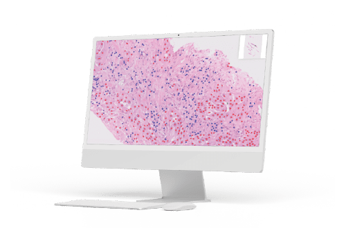
AI-Powered Image Structuring
Structure and characterize tissue and cell architecture from H&E, IHC, and Multiplex images.
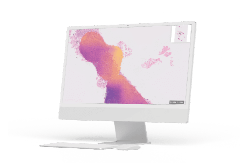
Spatial Feature Extraction
Calculate spatial features to better explain mechanism of action and biological phenomena.
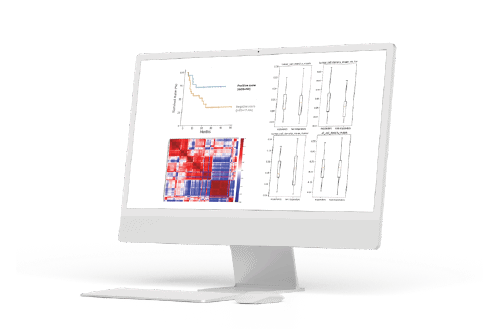
Predictive Models & Novel Insights
Leverage spatial analysis to predict outcomes, genomic status, and protein signatures.
Our Poster Presentations for SITC 2022
We are proud to present results from 3 studies at SITC this year. More details to follow.
Location: 1289
Predicting response to immune checkpoint inhibitors (ICI) in non-small-cell lung cancer (NSCLC) by combining spatial analysis of cells and RNA sequencing data from biopsies using deep learning (DL)
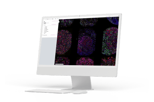
Nucleai ATOM offers powerful multiplex analysis. The platform is agnostic to staining and scanning platforms and is currently available in a comprehensive service mode, providing fast turnaround times for high plex, large datasets.
Image management and visualization: the viewer is designed to view high plex images with custom overlays, annotations, and algorithmic results