
Multiplex Imaging AI Platform for Large-Scale Academic Research
From multiplex images to reproducible spatial insights – built for large spatial cohorts
Nucleai’s multiplex imaging AI platform is an end-to-end, cloud-based analytics platform that transforms complex multiplex immunofluorescence (mIF) data into standardized, quantitative spatial insights. Designed for academic labs working at scale, the platform collapses the tissue-to-insight cycle from months to weeks, removing analytical bottlenecks that slow discovery.
*Submissions are reviewed to ensure alignment with study scale and workflow requirements.
Priority Access Program
Priority access for leading spatial biology research groups
Nucleai is opening priority access to leading academic and hospital-based research labs working with large mIF cohorts (50+ cases). This program is designed for groups looking to:
- Scale spatial analysis without fragmented tools or custom code
- Standardize analysis across large studies and collaborators
- Reduce time spent on pipeline maintenance and manual QC
Generate high-quality, reproducible results for publication
The challenge with mIF at Scale
Spatial imaging technologies have advanced rapidly, but downstream analytics remain a critical bottleneck. As mIF studies grow in size and dimensionality,
- Fragmented workflows force researchers to stitch together multiple tools across segmentation, phenotyping, QC, and spatial analysis
- High technical burden slows studies due to custom scripting and manual intervention
- Limited scalability makes large cohorts difficult to analyze consistently
- Reproducibility gaps emerge across studies and collaborators
The result: labs spend weeks or months managing data instead of advancing biological insight.
The Nucleai Solution
An AI-enabled, end-to-end mIF analytics platform built for scale
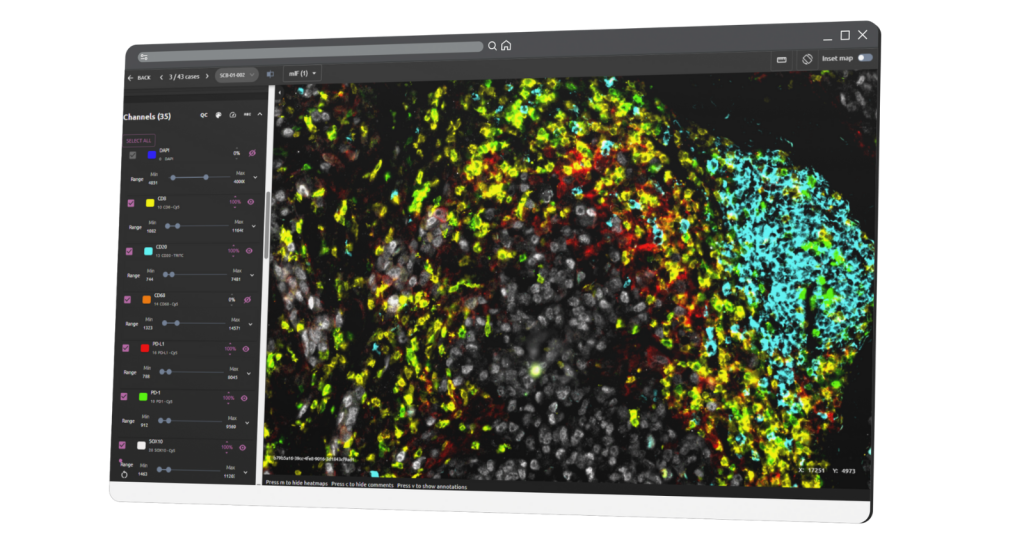
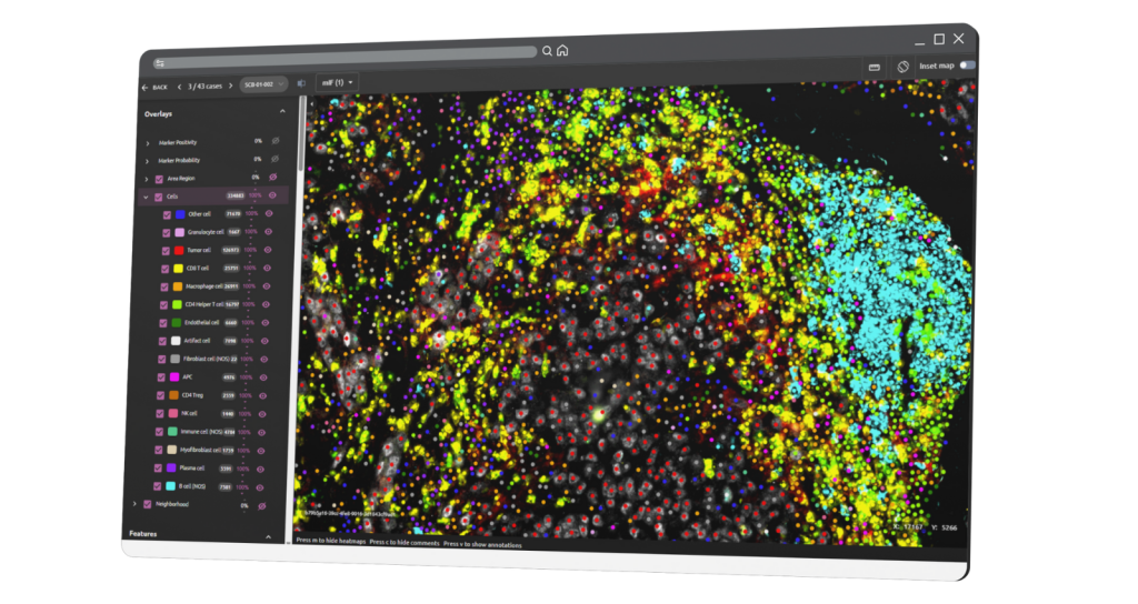
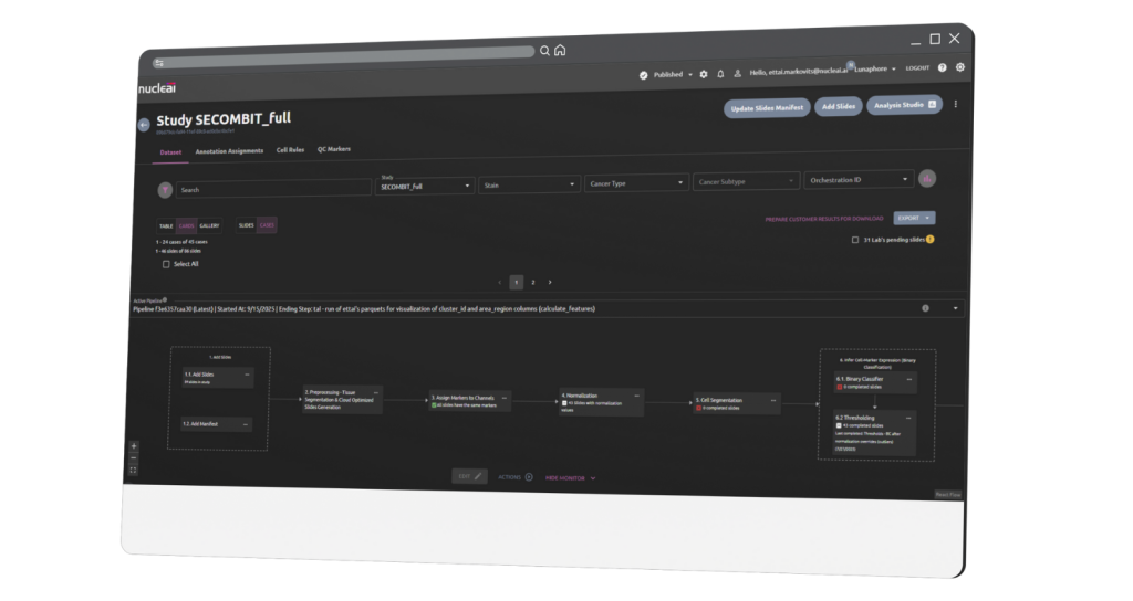
Nucleai’s Multiplex Imaging AI Platform unifies the full mIF analysis workflow in a single, cloud-native system – from ingest to structured outputs for downstream spatial analysis.
Core capabilities include:
- Automated ingest, manifest management, and study-level QC
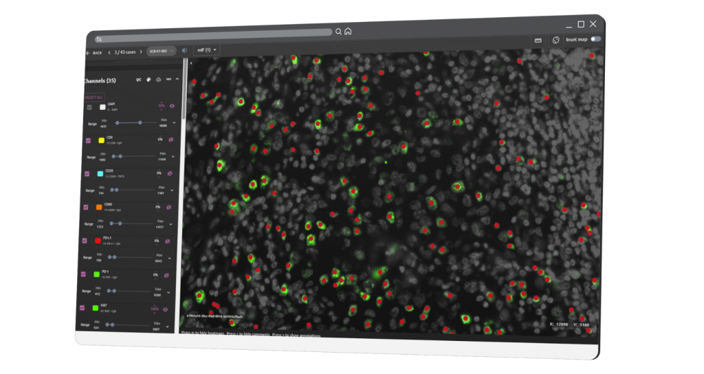
- Robust automated normalization and cell segmentation across multiple stainers
- AI-driven cell typing and marker positivity
- Standardized, exportable cell and image-level outputs
Designed to scale: The platform is designed to work across instruments and datasets, enabling consistent, repeatable analysis across hundreds of images without manual pipeline rework.
Key benefits of Nucleai platform
Compared to existing multiplex imaging software, Nucleai’s platform is optimized for large-scale studies where speed, consistency, and reproducibility matter most:
- Scalable workflows: Unified, automated pipeline from raw images to insights, enabling reliable performance in large cohorts.
- Reproducible analytics: Standardized outputs across studies and collaborators.
- Accelerated discovery: Automated workflows reduce tissue-to-insight timelines from months to weeks.
- Cloud-native SaaS: No local infrastructure, scripting, or fragmented tooling
By removing analytical friction, Nucleai enables more experiments, deeper biological insight, and faster translation from spatial data to discovery.
Request priority access
We are inviting a small number of leading academic labs to participate in priority access.
*Submissions are reviewed to ensure alignment with study scale and workflow requirements.