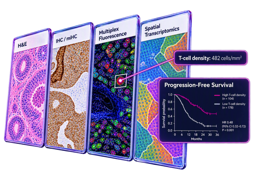
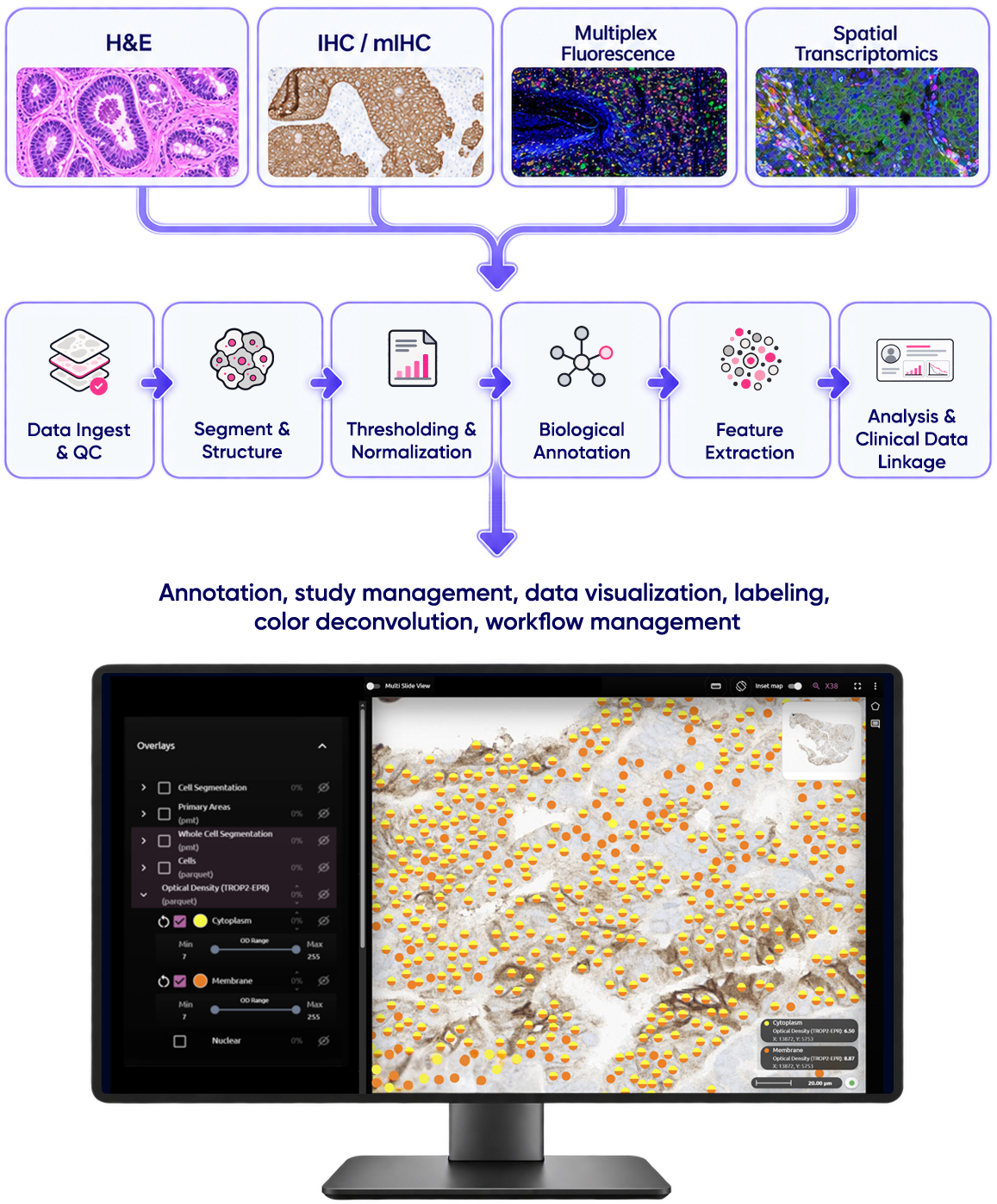
Nucleai’s tissue intelligence platform extracts structured, reproducible spatial features from H&E, IHC, multiplex imaging, and spatial transcriptomics modalities, then links those features to why a therapy works and which patients will benefit.
Most tissue analysis stops at percent positivity and an H-score — numbers that flatten everything happening on the slide. The signal that explains response, resistance, and survival lives elsewhere: how features are distributed across a slide, how they vary between disease sites, and how they differ from patient to patient.
Nucleai’s platform is built to read that complexity. Models adapt to the data in front of them rather than arriving pre-configured, extracting spatial features at a depth computationally assisted pathologists cannot reach alone. Those features are paired across sections and modalities so an entire cohort can be interrogated as one unified dataset.
Four Modalities, One Framework
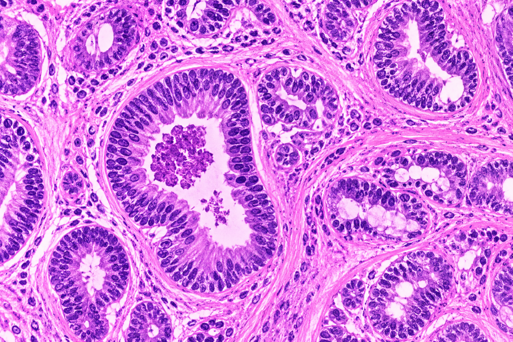
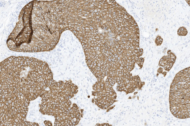
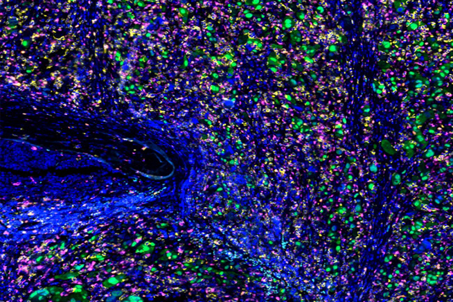
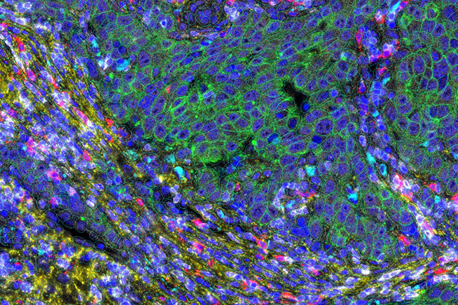
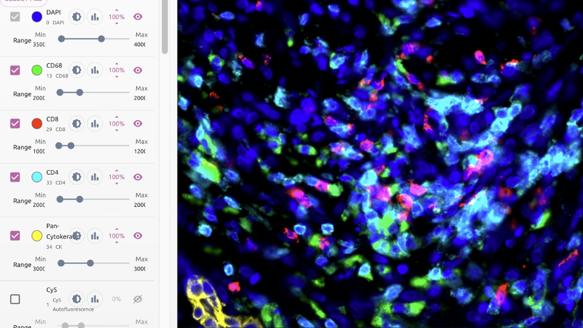
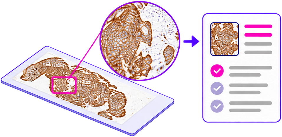
The platform applies one analytical framework across every major tissue data type: H&E for cell and tissue-region detection, IHC/mIHC for continuous per-cell marker scoring, multiplex fluorescence for generalizable cell typing, and spatial transcriptomics for multiomic integration — so findings stay comparable no matter which image produced them.
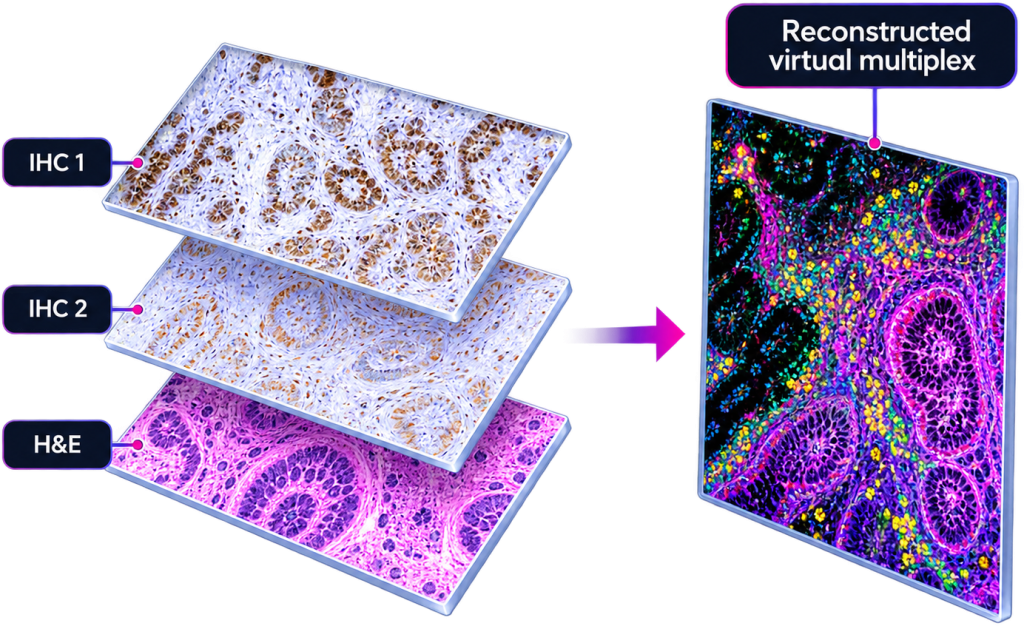
Additional capabilities push past what any single stain can show: virtual multiplexing, which registers serial H&E, IHC, and mIF sections into one reconstructed view, and cross-modal prediction, which infers higher-cost readouts directly from routine H&E.
Before analysis begins, automated QC evaluates every marker and slide, and per-channel normalization keeps results comparable across batches and sites.
Traceable by Design
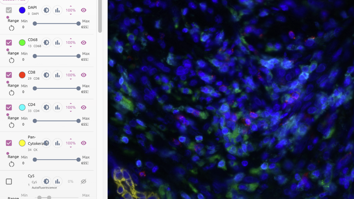
Every finding traces back to the image it came from, through a cross-modal viewer with downloadable overlays and reports. The platform is developed and deployed under design controls, supporting a validated path to CDx deployment through CLIA/CAP-certified partner labs.
Nucleai's products are designed, developed and deployed utilizing risk-based implementation of our ISO 13485 compliant quality management system as well as our ISO/IEC 27001 compliant information security management system.
Closing the Loop on Outcomes
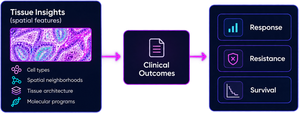
Spatial features are only as valuable as the clinical questions they answer. The platform can run inside your own environment, letting your team cross-reference Nucleai’s features against proprietary clinical endpoints, establishing which features actually explain response, resistance, and survival, without outcome data leaving your firewall.
Applied across 30+ clinical trials and 10+ indications.