
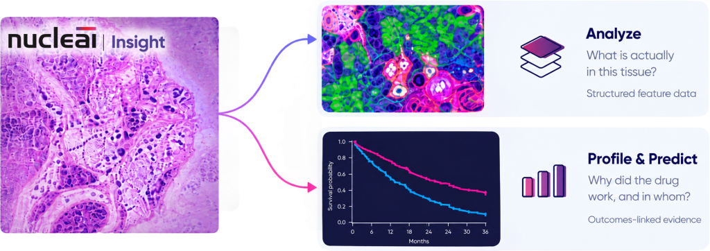
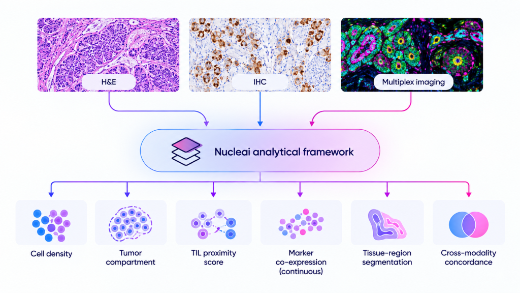

In a multi-site validation with Merck KGaA presented at USCAP 2023, a deep learning PD-L1 TPS scoring model was trained across five laboratories, three scanners, and two antibody clones on roughly 100,000 pathologist-annotated cells. It was then validated against two independent pathologists on an unseen 107-slide cohort at an R² of 0.91 against pathologist consensus. In a separate study presented at SITC 2023, pairing low-plex multiplex imaging with same-slide H&E reclassified six million previously marker-negative cells and identified tertiary lymphoid structures at 88% accuracy.

Translational pathologists and clinical biomarker leads scoping a first program engagement.

Nucleai can assess a delivered slide set on its own, flagging which slides will be excluded and why, and what that means for the analysis that is actually achievable. Sponsors routinely do not have this information about their own cohorts until after the fact.

Nucleai can shape the assay before staining, not analyzing after it. Nucleai works alongside sponsors and staining labs to build AI-readiness into panel design, set marker concentrations for quantitative rather than visual review, and catch weak dynamic range before it compromises cell calls downstream.
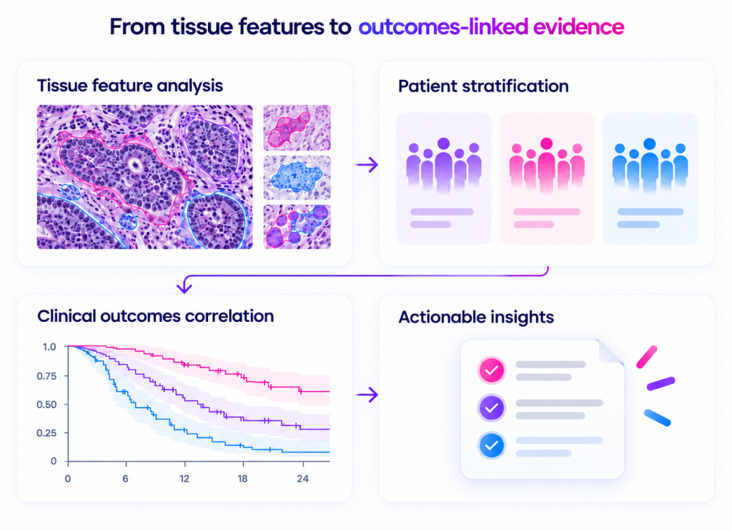
In a peer-reviewed collaboration with Adlai Nortye published in Cancers (2026), spatial features read from randomized Phase 2 H&E were associated with overall survival benefit.
Clinical biomarker leads, supported by a computational biology team.